

【研究成果】2016年-
研究成果92
Protein expression guided chemical profiling of living cells by the simultaneous observation of Raman scattering and anti-Stokes fluorescence emission
Liang-da Chiu, Taro Ichimura, Takumasa Sekiya, Hiroaki Machiyama,Tomonobu Watanabe, Hideaki Fujita, Takeaki Ozawa & Katsumasa Fujita
Scientific Reports 7, Article number: 43569 (2017)
Our current understanding of molecular biology provides a clear picture of how the genome, transcriptome and proteome regulate each other, but how the chemical environment of the cell plays a role in cellular regulation remains much to be studied. Here we show an imaging method using hybrid fluorescence-Raman microscopy that measures the chemical micro-environment associated with protein expression patterns in a living cell. Simultaneous detection of fluorescence and Raman signals, realised by spectrally separating the two modes through the single photon anti-Stokes fluorescence emission of fluorescent proteins, enables the accurate correlation of the chemical fingerprint of a specimen to its physiological state. Subsequent experiments revealed the slight chemical differences that enabled the chemical profiling of mouse embryonic stem cells with and without Oct4 expression. Furthermore, using the fluorescent probe as localisation guide, we successfully analysed the detailed chemical content of cell nucleus and Golgi body. The technique can be further applied to a wide range of biomedical studies for the better understanding of chemical events during biological processes.
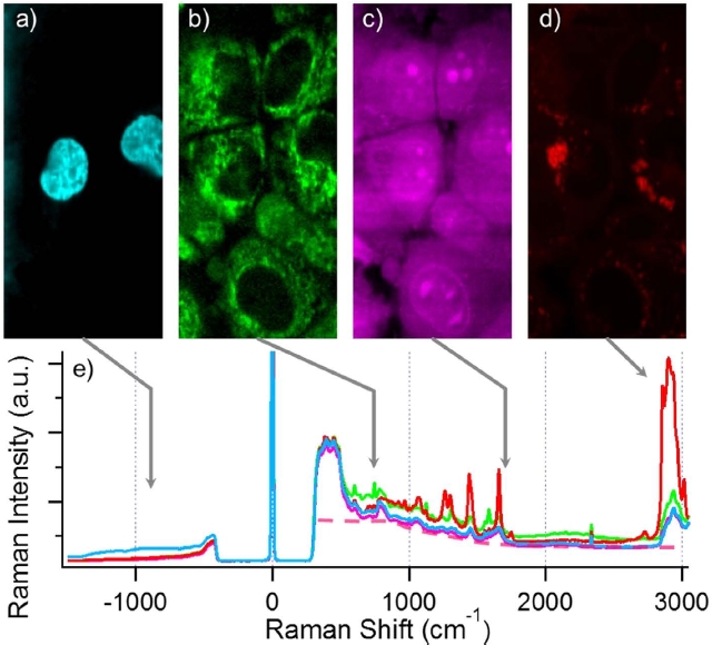
(a∼e) are the anti-Stokes fluorescence image (a), resonance enhanced 750 cm−1 cytochrome c Raman image (b), 1680 cm−1 amide I Raman image that shows protein contrast (c), 2852 cm−1 CH2 stretch Raman image that shows long chain lipids (d), and the hybrid spectrum taken from a random pixel of the corresponding contrasts (e). Cyan is the spectrum with strong CFP emission, green is with strong cytochrome c contrast, magenta from strong protein contrast and red from strong lipid contrast. Note that all these information are acquired within a single scan of the sample.