
【研究成果】2015年
研究成果89
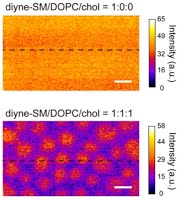
Sphingomyelin distribution in lipid rafts of artificial monolayer membranes visualized by Raman microscopy
Jun Ando, Masanao Kinoshita, Jin Cui, Hiroyuki Yamakoshi, Kosuke Dodo, Katsumasa Fujita, Michio Murata, and Mikiko Sodeoka
4558–4563| PNAS | April 14, 2015 | vol. 112
We synthesized an analog of sphingomyelin(SM) tagged with a small Raman active diyne moiety, which provides high chemical selectivity without affecting the membrane properties. Raman microscopy successfully visualized, at single lipid-layer sensitivity, a heterogeneous spatial distribution of this probe within raft-like ordered domains, which was different from the generally accepted raft model. This approach provides both chemical selectivity and quantitative imaging capability and is useful for functional studies of lipid rafts.