
【研究成果】2015年
研究成果85
Implementation of simultaneous quantitative phase with Raman imaging
Nicolas Pavillon and Nicholas I Smith
EPJ Techniques and Instrumentation (2015) 2:5
DOI 10.1140/epjti/s40485-015-0015-9
We present a technical overview of a multimodal system combining Raman microspectroscopy and quantitative phase microscopy (QPM), which allows two independent and simultaneous measurements of both the local molecular content and dynamic sample morphology. We present in detail the setup implementation and measurement procedure, and show how different features of QPM can be used to ensure optimal Raman measurement conditions and matched fields of view, through off-line calibration procedures such as digital propagation of the measured complex field and analysis of the system’s optical aberrations which can then be employed for numerical compensation and calibration. We present measurements on live cells, where images based both on the quantitative phase signal and on the Raman molecular contrast can simultaneously be retrieved and compared. The dynamic measurements obtained from QPM also enable the monitoring of the cell morphology during the laser scanning of the Raman measurement, making it possible to identify the movements which may occur during the measurement.
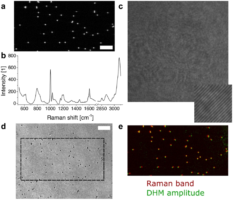
Multimodalmeasurement of polystyrene beads and calibration of the two imaging modes. The beads can be seen (a) in the Raman channel at 1003 cm−1, with (b) their corresponding spectrum. They can also be identified as defocused diffraction patterns in (c) the hologram, or as black dots in the corresponding (d) reconstructed amplitude image. (e) The images are then automatically registered, and shown merged with the Raman channel in red and the inverted DHM amplitude in green. Slight remaining discrepancies in position result from distortions between the two modes. Scale bars are 10 μm.
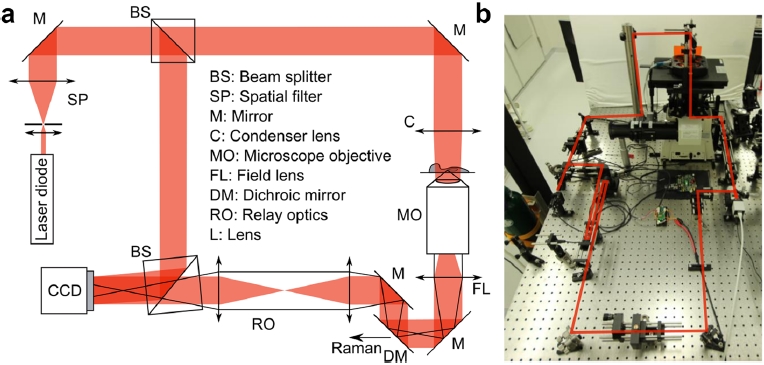
Schematic and implementation of the DHM setup


大阪大学フォトニクスセンター
Photonics Center, Osaka University