

【研究成果】2015年
研究成果80
Structured line illumination Raman microscopy
Kozue Watanabe, Almar F. Palonpon, Nicholas I. Smith, Liang-da Chiu, Atsushi Kasai, Hitoshi Hashimoto, Satoshi Kawata & Katsumasa Fujita
NATURE COMMUNICATIONS | 6:10095 | DOI: 10.1038/ncomms10095
Structured-line illumination (SLI) Raman microscopy that we have developed shows a well-balanced performance in spatial and spectral resolution with the capability of optical sectioning and powerful spectral analysis. The spatial resolution provided by SLI can reach the theoretical limit of confocal microscopy in many practical applications. Considering the SNR in the detection of Raman scattering, SLI Raman microscopy can realize a remarkable improvement in spatial resolution over that of current Raman imaging techniques, and which can further expand the application of Raman microscopy in various research fields such as material sciences, life sciences and pharmaceuticals. For example, our technique can lead to the detailed analysis of graphene, which is important in understanding fundamental questions on graphene sheet growth mechanism and characterization of graphene devices. In cell biology, as fluorescence SIM has contributed to visualize the detailed structure of cellular components, it might also be possible to spectrally distinguish these components even without staining by using the SLI Raman technique. In pharmaceuticals, an emerging demand that can benefit from the high spatial resolution of SLI Raman microscopy is the precise determination of the distribution of the different chemical components in a drug.
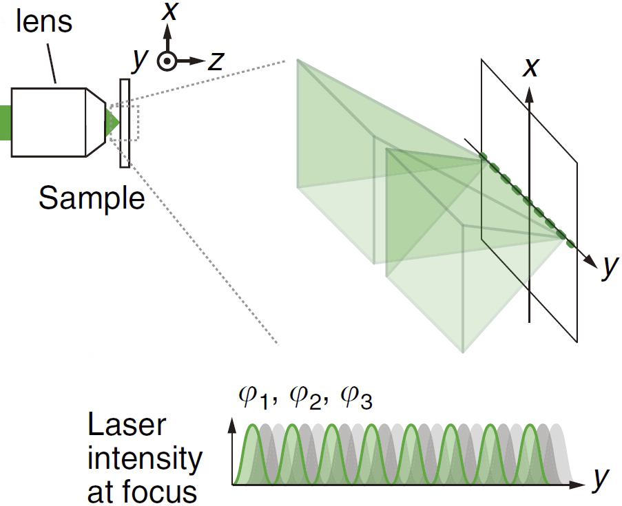
Light from the laser is split by a phase grating to produce interference fringes in the line illumination at the sample.
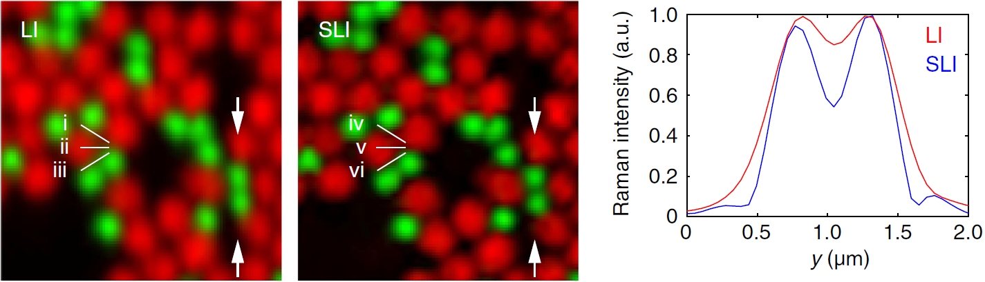
Raman images of a mouse brain slice. (a,b) The intensity distribution of Raman peaks at 1,682 cm-1 (red) and 2,848 cm-1 (green) imaged by the LI (a) and SLI (b) microscopes (Scale bar, 5 um). Raman peaks at 1,682 cm-1 and 2,848 cm-1 can be assigned to amide-I and CH2 stretching vibrational modes, respectively, predominantly observed in protein beta sheets and lipids.