

【研究成果】2012
研究成果43
Label-free Raman observation of cytochrome c dynamics during apoptosis
Masaya Okada, Nicholas Isaac Smith, Almar Flotildes Palonpon,
Hiromi Endo, Satoshi Kawata, Mikiko Sodeoka, and Katsumasa
Fujita
PNAS January 3, 2012 vol. 109, p. 28-32.
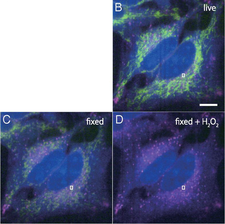
(B) Raman images of living HeLa cells, and the same cells were observed after (C) the paraformaldehyde fixation and (D) the H2O2 treatment following the fixation. For each image, Raman intensities at 750, 1686, and 2857 cm-1, which can be assigned to cytochrome c, Amide-I, and CH2 stretching vibration mode, are mapped in green, blue, and red, respectively. Scale bar, 10 μm.
We performed label-free observation ofmolecular dynamics in apoptotic cells by Raman microscopy. Dynamic changes in cytochrome c distribution at the Raman band of 750 cm-1, which is assigned to pyrrole breathing mode ν15 in cytochrome c, were observed after adding an apoptosis inducer to the cells. The comparison of mitochondria fluorescence images and Raman images of cytochrome c confirmed that changes in cytochrome c distribution can be distinguished as release of cytochrome c from mitochondria. Our observation also revealed that the redox state of cytochrome c was maintained during the release from the mitochondria. Monitoring
mitochondrial membrane potential with JC-1 dye confirmed that
the observed cytochrome c release was associated with apoptosis.