【研究成果】2012
研究成果41
High-speed molecular spectral imaging of tissue with stimulated Raman scattering
Yasuyuki Ozeki, Wataru Umemura, Yoichi Otsuka, Shuya Satoh, Hiroyuki Hashimoto, Kazuhiko Sumimura, Norihiko Nishizawa, Kiichi Fukui and Kazuyoshi Itoh
NATURE PHOTONICS VOL 6 |DECEMBER 2012
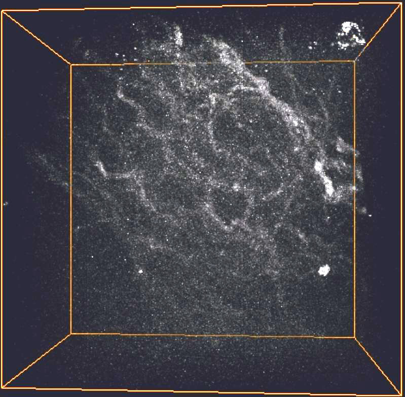
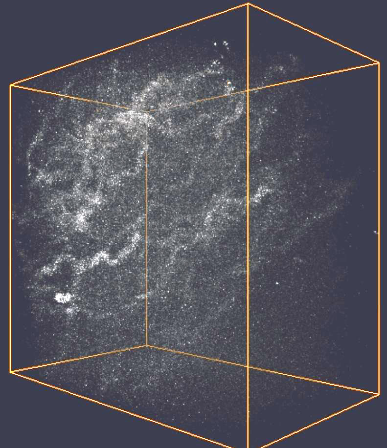
Three-dimensional, two-colour imaging of a vessel in rat liver. Images at wavenumbers of 2,850 and 2,950 cm-1 were successively taken while the z-position was scanned over 80 um in steps of 1 um. Total acquisition time is 5.3 s. Reconstructed three-dimensional image, Scale bars, 20 um.
We have demonstrated molecular spectral imaging based on high speed SRS (Raman scattering microscopy) spectral microscopy and modified ICA (independent component analysis). This system allows quick image acquisition at.30 frames/s by changing the wavenumber in a frame-by-frame manner. The modified ICA enables blind separation of constituents without a priori spectral data. The IC spectra give spectroscopic information, and the IC images can be used to produce multi colour images. This label-free and rapid observation method would be especially useful in medical imaging. We present various imaging modalities such as two-dimensional spectral imaging of rat liver, two-colour three-dimensional imaging of a vessel in rat liver, spectral imaging of several sections of intestinal villi in mouse, and in vivo spectral imaging of mouse ear skin.
In SRS microscopy, we use two-color laser pulses, one of which is intensity modulated in time. Then they are focused on a sample. When the optical frequency difference of the pulses matches the vibrational frequency of the sample molecules, the optical energy of the high-frequency pulse is transferred to the low-frequency pulse and to molecular vibrations through the SRS process. As a result, the intensity modulation is transferred to the other pulse, and this modulation transfer is measured by the lock-in detection technique. SRS has various advantages, such as accessibility to the vibrational spectrum without spectral distortion, quantitative image contrast, high sensitivity, and high imaging speed.
In SRS microscopy, we use two-color laser pulses, one of which is intensity modulated in time. Then they are focused on a sample. When the optical frequency difference of the pulses matches the vibrational frequency of the sample molecules, the optical energy of the high-frequency pulse is transferred to the low-frequency pulse and to molecular vibrations through the SRS process. As a result, the intensity modulation is transferred to the other pulse, and this modulation transfer is measured by the lock-in detection technique. SRS has various advantages, such as accessibility to the vibrational spectrum without spectral distortion, quantitative image contrast, high sensitivity, and high imaging speed.


大阪大学フォトニクスセンター
Photonics Center, Osaka University