

【研究成果】2016年-
研究成果95
Saturated two-photon excitation fluorescence microscopy with core-ring illumination
Ryosuke Oketani, Atsushi Doi, Nicholas I. Smith, Yasunori Nawa, Satoshi Kawata, and Katsumasa Fujita
Vol. 42, No. 3 / February 1 2017 / Optics Letters571
We demonstrated resolution improvement in two-photon excitation microscopy by combining saturated excitation (SAX) of fluorescence and pupil manipulation. We theoretically estimated the resolution improvement and the sidelobe effect in the point spread function with various pupil designs and found that the combination of SAX and core-ring illumination can effectively enhance the spatial resolution in 3D and suppress sidelobe artifacts. The experimental demonstration shows that the proposed technique is effective for observation with a depth of 100 μm in a tissue phantom and can be applied to 3D observations of tissue samples with higher spatial resolution than conventional two-photon excitation microscopy.
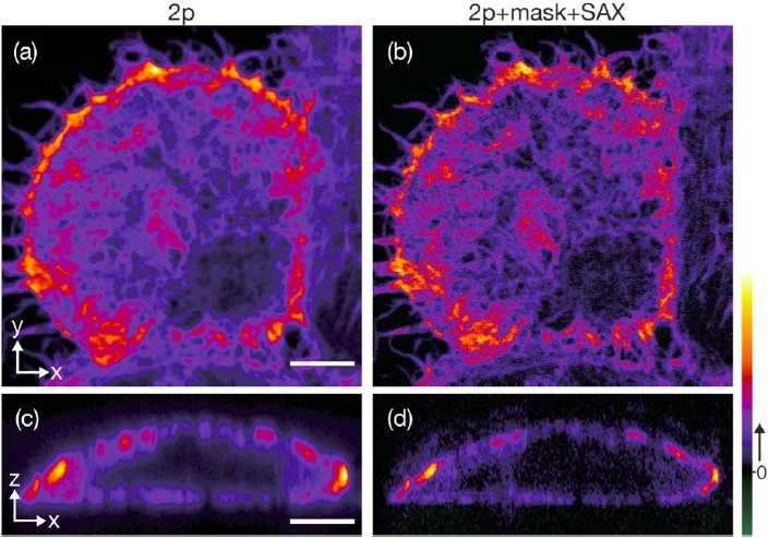
Images of filamentous structures of actin in fixed HeLa cells obtained with standard two-photon excitation microscopy [(a) and (c)] and two-photon SAX with core-ring illumination [(b) and (d)]. The scale bar is 5 μm. The excitation powers at the focus for unsaturated excitation with and without the mask were 2.0 and 1.1 mW, respectively. The exposure time was 200 μs for observations without SAX. The excitation power of 28.6 mW and the exposure time of 1 μs were used for SAX imaging with the mask. No image processing was applied.