

【研究成果】2012
研究成果42
Detection of influenza virus using a lateral flow immunoassay for amplified DNA by a microfluidic RT-PCR chip
Naoki Nagatani, Keiichiro Yamanaka, Hiromi Ushijima, Ritsuko Koketsu, Tadahiro Sasaki, Kazuyoshi Ikuta, Masato Saito, Toshiro Miyahara and Eiichi Tamiya
Analyst, 2012, 137, 3422
Influenza virus RNA was amplified by a continuous-flow polydimethylsiloxane microfluidic RT-PCR chip within 15–20 min. The amplified influenza virus RNA was observed with the naked eye, as the red color at the test line, using a lateral flow immunoassay within 1 min. The amplified DNA from influenza virus RNA, using a fluorescein isothiocyanate (FITC) labelled primer and biotinylated primer, by the microfluidic continuous-flow reverse transcription PCR (RT-PCR) chip was detected by amplified DNA detection lateral flow immunoassay (ADLFIA). The sensitivity of ADLFIA is comparable to that of agarose gel electrophoresis. The detection by ADLIFA needs only the naked eye. ADLFIA will be appended to a conjugate pad containing a dried excess volume of biotin antibody-labelled gold nanoparticles for a one-step assay. The conjugate pad appending ADLFIA can integrate easily into the solution exuding from the outlet of the chip. The integration enables the easy detection by ADLFIA without a mixing step. We are continuing our effort towards a battery powered portable PCR chip, and a battery powered portable PCR chip with ADLFIA has the potential to give the portable system a diagnostic test.
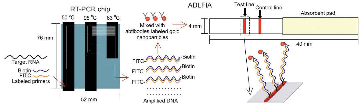
Schematic representation of the detection of the influenza virus using the continuous-flow RT-PCR chip and ADLFIA. Influenza virus RNA (target RNA) is amplified by the continuous-flow RT-PCR chip (52 x 76 mm2) using an FITC labelled and biotinylated primer set. The amplified DNA is mixed with biotin antibodies labelled with gold nanoparticles. The size of the ADLFIA strip is 4 x 40 mm2. FITC antibody and mouse IgG antibody are immobilized on the test and control line, respectively. The mixture is absorbed for ADLFIA, the amplified DNA complex with biotin antibody-labelled gold nanoparticles is captured by the FITC antibody at the test line. Finally, a red color appears as the result of accumulation of gold nanoparticles.